
Overview
Electroporation
Founded in 1963, our laboratory has evolved from functional electrical stimulation research to become a world-leading center for electroporation science, spanning molecular mechanisms to clinical applications.
The beginnings
Since its foundation in 1963 by Professor Lojze Vodovnik, the Laboratory of Biocybernetics has been involved in the study of interactions between electromagnetic fields (EMFs) and biological systems. This includes the general investigation of effects of EMFs on organisms, as well as exploitation of EMFs for therapeutic and diagnostic purposes.
Until the end of the 1970s, our main research topic was functional electrical stimulation (FES) for the restoration of motor functions impaired by different types of injuries and neuromuscular diseases. This approach has since been adopted by many rehabilitation centers worldwide, and continues to be developed at our faculty in the Laboratory of Robotics in collaboration with the University Rehabilitation Institute.
Ongoing research
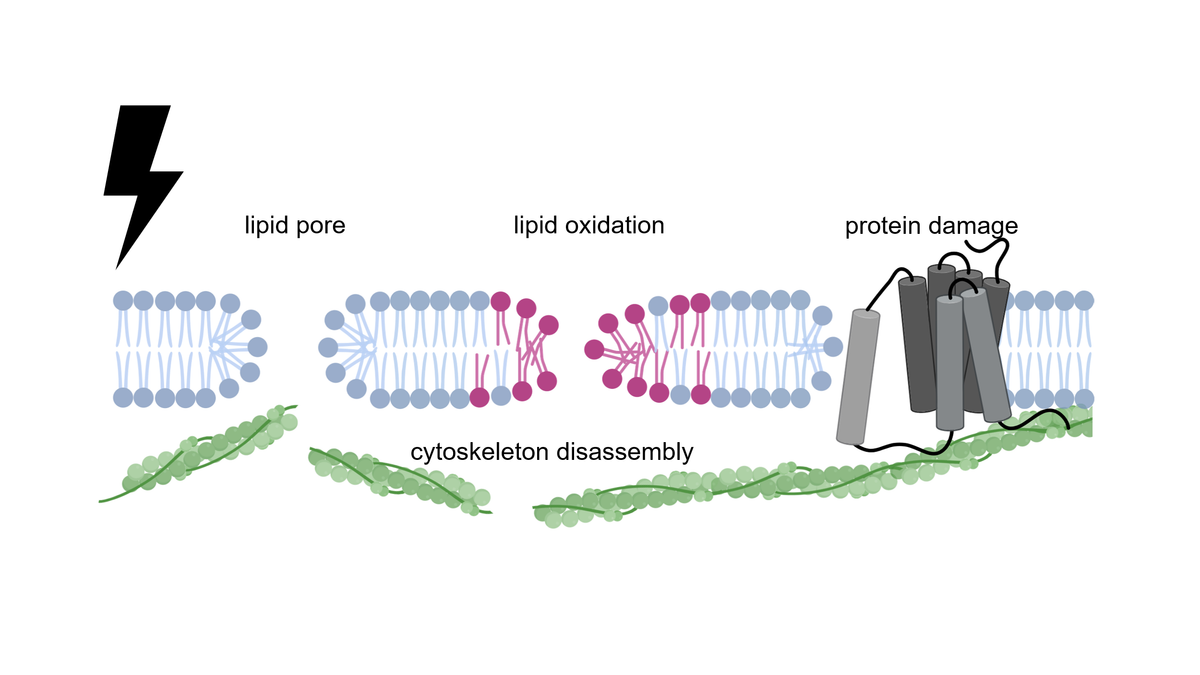
Since the 1980s, our focus gradually shifted to the influence of electric currents and electromagnetic fields on the physiological state of cells and tissues, particularly on cell membrane electroporation with its applications in biology, biotechnology, and medicine 1,2. While formation of electrically-induced aqueous pores in the lipid bilayer is now a widely recognized mechanism of this phenomenon, we now know that oxidative changes to individual membrane lipids and damage to membrane proteins also contribute to electroporation 3.
We study the electroporation phenomenon using both theoretical and experimental approaches, spanning scales from atomic-molecular (MD simulations), membrane-level (lipid bilayers and vesicles), organelle- and cell-level (cells in suspension, attached, and in 3D cultures), up to tissues and organs (animal studies in cooperation with the Veterinary Faculty of the University of Ljubljana and Sunnybrook Research Institute in Toronto, Canada). We also collaborate with medical institutions (Departments of Cardiovascular Surgery and Cardiology at the University Medical Centre Ljubljana, and Institute of Oncology Ljubljana) in clinical studies, and with national and international industrial partners in development of medical and experimental devices. Our work encompasses development of devices, fundamental and translational experiments and modeling, as well as treatment planning and development of information technology for clinical trials, and ranges from the studies of basic mechanisms to applications in oncology, cardiology, gene and cell therapy, as well as food processing and biotechnology. An organizational chart of the areas of our work is sketched below, while more detailed descriptions of our work in each field are available by following the links on the tab "Research".
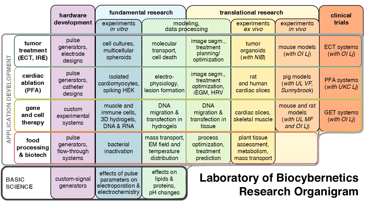
Our publications on electroporation and its applications have so far received over 30 thousand citations. Since 2003 we have been organizing (first bi-annually, since 2011 annually) the workshop and postgraduate course Electroporation-Based Technologies and Treatments (EBTT), attracting each year over 50 attendees, and in total over 800 participants from over 40 countries. We were the chairs of the European network for development of electroporation-based technologies and treatments – COST Action TD1104 (2011-2016) connecting 581 researchers of electroporation from 243 research institutions and 28 companies from 43 countries. Professor Damijan Miklavčič, the research head of the laboratory, is the editor of the Handbook of Electroporation published by Springer, with the first edition released in 2017 spanning almost 3000 pages, and the second, updated and extended edition slated for publication in 2027.
Molecular mechanisms of electroporation
The intense electric field in the membrane disrupts the lipid bilayer structure, and can also chemically modify membrane lipids and proteins. This creates new pathways for transport of ions and molecules across the membrane. At the molecular level, electroporation involves several mechanisms that can occur simultaneously. These include the formation of transient (metastable) aqueous pores in the lipid bilayer, oxidation of membrane lipids, and damage to certain membrane proteins, particularly ion channels and transporters. Both lipid oxidation and membrane protein damage can also lead to pore formation. Although these pores cannot be directly visualized with conventional microscopy due to their small size and metastability, their existence has been confirmed by molecular dynamics simulations and indirect experimental evidence. Furthermore, electroporation leads to changes in the cytoskeleton – the structural network inside the cell that is mechanically connected to the membrane and influences resealing kinetics. Together, these molecular-level changes determine the membrane permeabilization and subsequent cellular response. 3
Electroporation at the tissue level
When electroporation is performed in tissues, it is more complex than in isolated cells. The mutual proximity of cells and their intercellular connections significantly affect the distribution of the transmembrane voltage induced by the exposure to the electric field. Molecular transport through tissue is hindered due to tight cellular packing and the extracellular matrix surrounding them. The electric field distribution in tissue is inherently nonhomogeneous due to complex geometry and differences in electrical conductivity between tissue components. Moreover, electroporation dynamically changes tissue electrical properties during pulse delivery: electroporated regions become more conductive, which redistributes the electric field during pulse delivery. Understanding these interactions requires computational modeling that accounts for tissue heterogeneity, dynamic conductivity changes, and the interplay between field distribution and biological response.
Effects on cellular electrophysiology
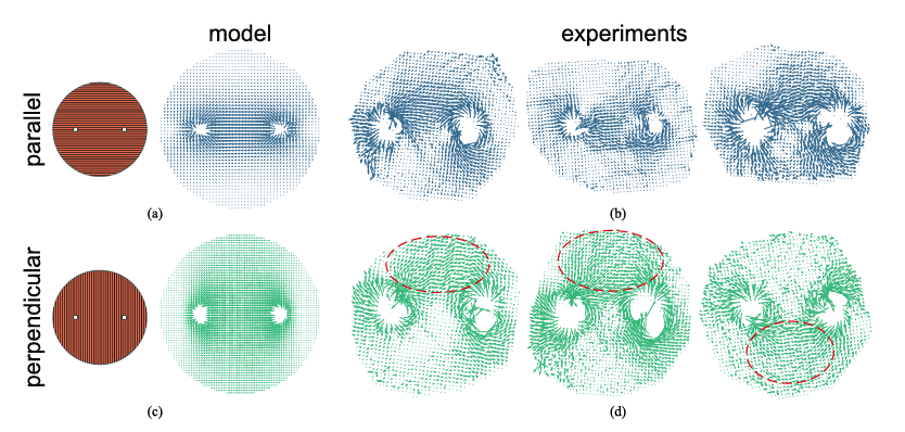
Low-voltage electric pulses can stimulate excitable cells and are routinely used in medical applications such as nerve stimulation and cardiac pacing. However, electroporation profoundly affects excitability in nerve and muscle cells. When electroporation occurs, additional nonselective currents flow through electroporated regions, which disrupts the normal cell signaling. Even mild electroporation can affect action potential generation and propagation, while stronger electroporation causes sustained membrane depolarization (stunning). In muscle cells, such as cardiomyocytes, electroporation also affects contraction. Additionally, electroporation impacts the electrophysiology of non-excitable cells and triggers intracellular calcium responses through both membrane permeabilization and ion channel activation. These responses can influence cell proliferation, differentiation, and cell death pathways, which is important to consider when designing electroporation-based treatments. 3
Applications in medicine and biology
Electroporation has been the main research area of the Laboratory of Biocybernetics since the 1990s. Over the years the field has matured in terms of our understanding of physical and biological phenomena involved in its complex and multifaceted effects on living cells, tissues, and the whole organism. Electroporation can be used nondestructively (reversible electroporation) to enhance or facilitate the transport of various substances across the cell membrane, or destructively as a direct trigger for loss of cell viability (irreversible electroporation). These effects can be exploited in very diverse applications. In the beginning, our research and development was primarily focused on applications of electroporation for treatment of solid tumors. We were one of the pioneers in the development of electrochemotherapy of tumors, in which reversible electroporation is used to enhance the effectiveness of chemotherapeutic drugs. Another use of reversible electroporation is gene electrotransfer (GET), in which electroporation is used to introduce genetic material into cells to be exploited for gene and cell therapy. On the other hand, irreversible electroporation is used for ablation (destruction or removal of abnormally functioning tissue). The so-called pulsed field ablation (PFA) can be used, for example, for destruction of tumors and in treatment of cardiac arrhythmias. Over the last decade our attention has gradually shifted to application of electroporation in cardiology to help develop new approaches to treat life-threatening arrhythmias, most notably for treatment of atrial fibrillation, in which PFA is already overshadowing the conventional thermal ablation methods. The exciting story of electroporation applications is far from over, as the existing treatments need to be refined, and new target tissues and diseases to be treated are on the horizon.