Modelling of electric current and electromagnetic field distribution gives valuable insight into the process of electroporation. Experimenting on models is easier and much more flexible than on real biological systems, and sometimes it is also the only way to evaluate the role of certain stimulation conditions. Still, one should keep in mind that a model is a rather simplified version of real conditions. As such, it provides additional information and help in planning of experiments, but it can not substitute studies on biological systems.
Two distinct approaches are used in modelling – the analytical and the numerical one. The analytical methods are physically more revealing, yielding solutions is form of expressions, but their range of applicability is limited to simple geometries and linear phenomena. In contrast, the numerical approach can be applied to very intricate conditions, but fails to give explicit relations. The heterogeneous composition of tissues makes the analytical approach practically impossible, but numerical methods are suitable, especially the finite elements method. The essence of this method is the division of the model into small units (finite elements), inside which the electrical properties follow a defined function, and then solving the equations for each of these units. For this purpose we are equipped with the state of the art computer workstations and software for computer modelling such as COMSOL Multiphysics (by Comsol Inc.).
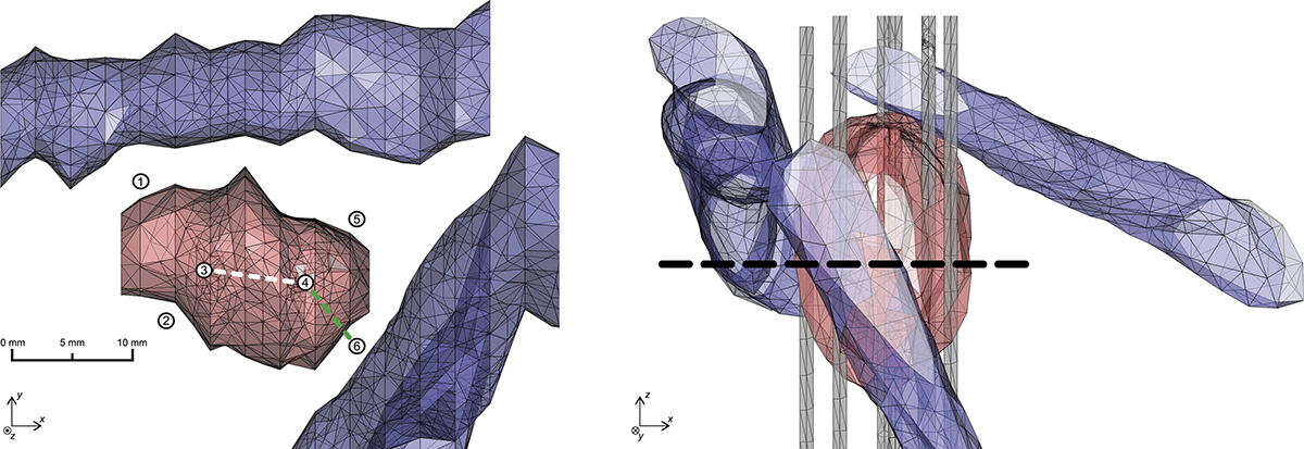
Figure: A 3-D numerical model of a deep-seated tumor in a liver with inserted needle electrodes. The tumor (in red) was located between the inferior vena cava and main hepatic veins (all in blue). Ref: Kranjc et al, PLOS One, 2012.